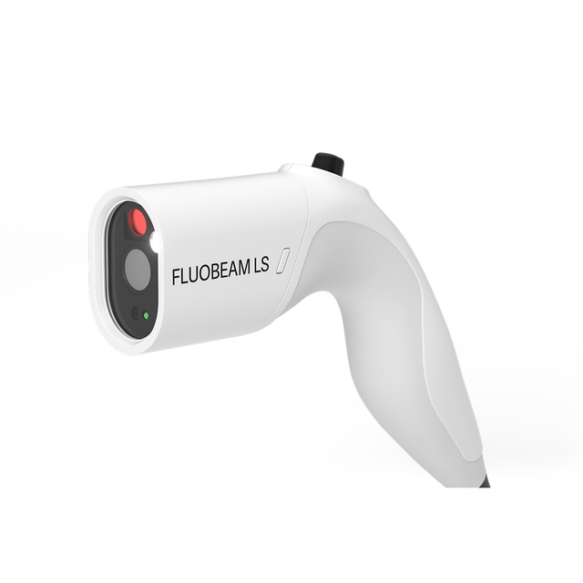
Supporting surgeons when it matters
Designed to meet the needs of the operating room, FLUOBEAM LS redefines fluorescence imaging by combining advanced ergonomics, high definition image quality, and clinical decision‑support tools. It assists surgeons in identifying and preserving critical anatomical structures, supporting intraoperative decision.
Explore Fluobeam LS
Explore Fluobeam LS
Removable cable from control box
with its direct access to software
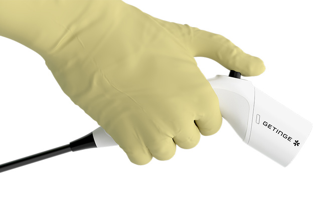
Optimized ergonomics supporting surgical procedure
The ergonomics of FLUOBEAM LS has been optimized to provide:
- Greater ease of use
- Unobstructed surgical field
- Seamless integration into surgical workflows
Productos relacionados
Compact design for effortless handling
The ergonomics of FLUOBEAM LS have been redesigned to offer surgeons intuitive and lightweight handling throughout the procedure. Its compact and optimized design makes it a useful tool for interventions requiring small incisions, supporting precise manipulation and comfortable use.
- Lightweight, intuitive handling
- Compact design suited for small‑incision procedures
- Comfortable use throughout the procedure
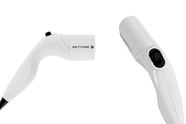
Keep control
The integrated joystick allows surgeons to directly control key system functions from within the sterile field.
- Uninterrupted surgical workflow
- Reduced need for external interactions
- Intuitive navigation in user interface
The size and the working distance make the difference
Optimized for short working distances, FLUOBEAM LS is designed to explore deep tissues within incisions. Moreover, its compact design allows direct visualization of the surgical field within the incision, without obstructing the surgeon’s line of sight.
- Positioning very close to the incision
- No obstruction of the surgeon’s direct field of view
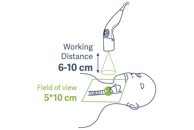
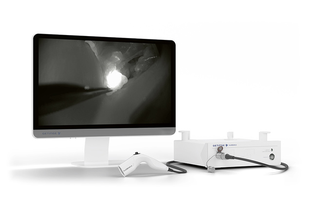
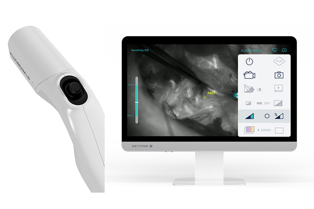
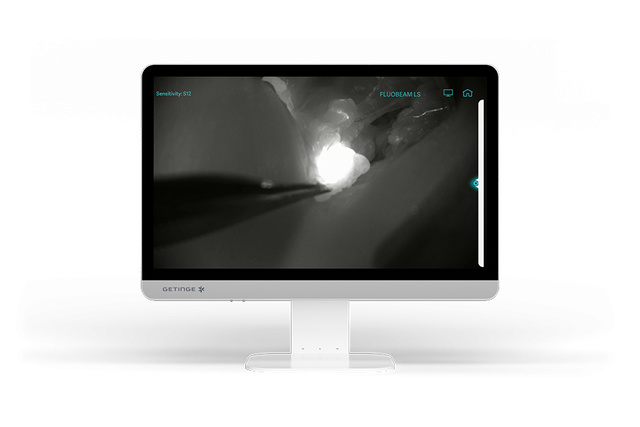
Image quality serving clinical expectations
FLUOBEAM LS is equipped with a high‑resolution imaging camera essential for detailed visualization of small anatomical structures such as blood vessels. The resulting image quality provides enhanced contrast and precision, supporting clearer intraoperative interpretation, particularly during delicate surgical procedures.
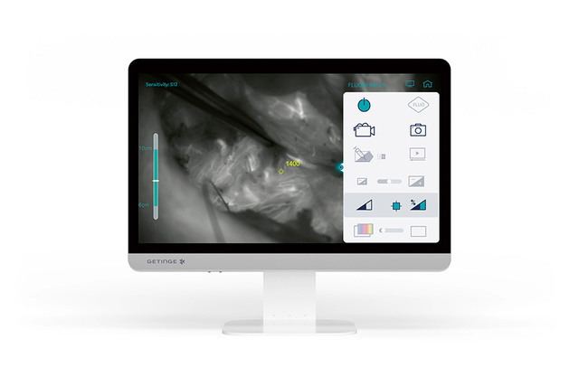
Absolute and Relative Quantification
The quantification tool provides objective intraoperative information on ICG perfusion through vascular or lymphatic network. The information is provided in both absolute and relative values. This objective data supports the surgeon’s assessment.
Precision, simplicity and innovation in surgical imaging
In surgery, the identification, preservation and assessment of critical anatomical structures represent a major challenge. Some structures remain difficult to distinguish with the naked eye, even for experienced surgeons, due to their small size, deep location or anatomical variability.
In this context, intraoperative fluorescence imaging has become an essential tool to enhance visualization and support decision making during surgery. FLUOBEAM LS delivers user experience, combining intuitive handling with precise, real‑time information that empowers surgeons throughout every step of the procedure.
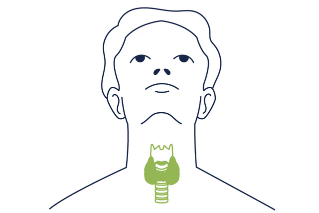
FLUOBEAM LS: fluorescence imaging serving thyroid surgery
Compact and intuitive, FLUOBEAM LS provides optimal tissue visualization. Because preserving the parathyroid glands and their vascularization is essential to reducing the risk of postoperative hypocalcemia, the system offers surgeons real‑time and objective support throughout the procedure.[1] Its high‑resolution camera and integrated quantification tool enhance safety and confidence during delicate steps.
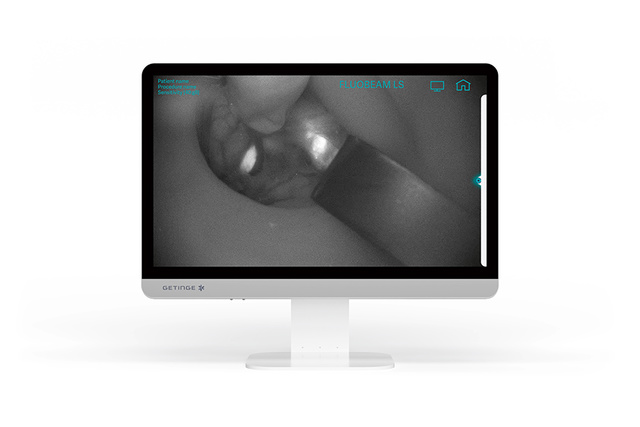
Parathyroid gland detection by autofluorescence
FLUOBEAM LS enables real-time visualization of parathyroid glands without the need for a contrast agent, leveraging their natural autofluorescence throughout the dissection. This capability allows for early and reliable detection of the parathyroid glands during thyroidectomy or lobectomy, supporting safer surgical decision-making.[2] [3]
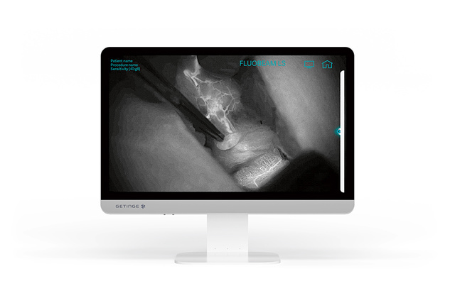
ICG fluorescence imaging for parathyroid vascular assessment
Indocyanine green (ICG) fluorescence imaging enables precise assessment of parathyroid gland vascularization by visualizing vascular pedicles and evaluating vascular dynamics in real time during surgery. At the end of the procedure, surgeons can verify in situ the viability of the parathyroid glands.
FLUOBEAM LS also provides quantitative measurements of the fluorescence signal after ICG injection, reflecting the perfusion status of each gland. This includes both relative and absolute assessment of vascularization levels, ensuring objective confirmation of visual information. Together, these capabilities support more reliable decision-making during delicate endocrine surgeries.
Quantification Tool: keeping you in control throughout surgery
The quantification tool provides intraoperative information on the perfusion level of the parathyroid glands. It integrates easily into the surgical workflow. Once the preliminary level of dissection is reached, the surgeon identifies the parathyroid gland using autofluorescence. After completing the dissection, the surgeon can then assess the perfusion of the parathyroid glands. At this stage, the quantification tool provides objective information expressed as absolute or relative values on the quality of parathyroid gland perfusion.
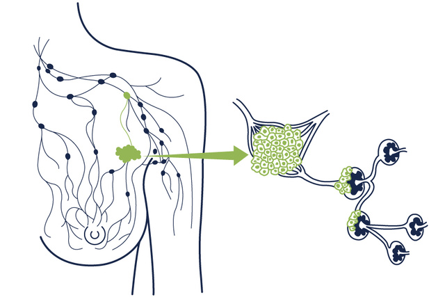
Axillary sentinel lymph node biopsy
Fluorescence imaging with Indocyanine Green (ICG) has recently emerged as a valuable alternative for surgical guidance in sentinel lymph node (SLN) detection.[4] Designed to meet the specific constraints of SLN biopsy, FLUOBEAM LS is a compact, lightweight and user‑friendly device optimized for use close to the incision. Its ergonomics and intuitive handling offer a reliable solution that integrates smoothly into the surgical workflow and helps improve the patient care pathway.
The device provides clear visualization of superficial lymphatic drainage and facilitates sentinel lymph node detection after incision across various tumor types, offering surgeons a precise and efficient guidance tool.
Superficial lymphatic drainage visualization for sentinel lymph node detection
Fluorescence imaging with Indocyanine Green (ICG) offers a complementary approach for sentinel lymph node (SLN) detection. It provides high clinical performance with detection rates reported as comparable to established techniques. FLUOBEAM LS offers direct and real‑time visualization, enabling surgeons to clearly follow superficial lymphatic drainage and identify the sentinel lymph node during biopsy. Its high‑resolution imaging provides sharp, detailed views that enhance the accuracy of surgical decision‑making.
The workflow is also simplified, as the procedure typically requires only a single ICG injection performed in the operating room on the day of surgery. Designed for ease of use in the operating room, the device features a quick learning curve and an intuitive interface that supports fast adoption.
This streamlined process contributes to surgical efficiency and patient comfort, making fluorescence an easy‑to‑use tool for daily practice.
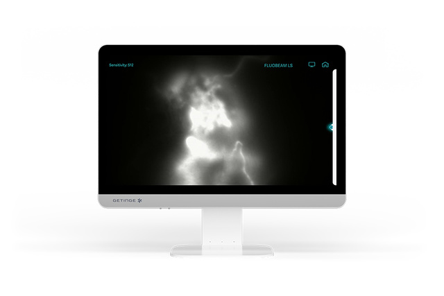
Superficial lymphatic drainage visualization
Detection of sentinel lymph node after skin incision
Key benefits
- Miniaturization of the optical head allowing visualization at the incision
- Optimized working distance, enabling in-depth exploration of incisions for localization of the sentinel lymph node
- Full HD image quality providing detailed visualization of small anatomical structures
Marketing Sales - Brochures
-
Brochure describing the Fluobeam LS, a fluorescence imaging camera designed for the identification of parathyroid glands and the detection of lymph nodes.