No two breaths are the same
Deep or shallow, long or short, with larger or smaller volumes – that is how we breathe naturally. Conventional intensive care ventilation cannot fully replicate this variability. With Getinge’s pioneering NAVA ventilation method (NAVA = Neurally Adjusted Ventilatory Assist), patients can control their own ventilation patterns neurally, allowing a more physiological and natural form of mechanical ventilation.
The patient controls the ventilation
Natural breathing is initiated by the respiratory center in the brain and mainly accomplished by the diaphragm, the main muscle responsible for breathing. When the diaphragm contracts, we inhale; when it relaxes, we exhale. This is where the concept of neurally controlled ventilation support comes into play.
NAVA measures the patient’s respiratory impulses using an Edi catheter, which is inserted into the stomach and positioned near the diaphragm. The electrodes on the catheter measure the electrical activity of the diaphragm and send a real-time signal indicating that the patient wants to breathe. The ventilator then delivers air and oxygen in synchrony with this Edi signal. When the electrodes detect that the diaphragm’s activity has ended, the ventilator allows exhalation.
NAVA is thus a ventilation mode in which both the timing and the level of support are controlled neurally by the patient. It is not the ventilator but primarily the patient who controls the ventilation: the initiation of each breath, tidal volume, and termination. The result is improved synchrony between patient and ventilator – in both invasive NAVA and non-invasive NAVA (NIV NAVA).
No longer just breathing for the patient, but with the patient
Patients who are connected to a ventilator usually have a functioning diaphragm initially, but it quickly weakens when the ventilator begins to breathe for them. Conventional ventilation modes do not monitor diaphragmatic activity, so the full effects of mechanical ventilation on the diaphragm remain unclear. For example, if the applied pressure support is too high, the patient’s own respiratory drive is suppressed, and the diaphragm becomes inactive and weakened. Over time, this can make it difficult to wean the patient from the ventilator.
With NAVA, the electrical activity of the diaphragm is continuously monitored, ensuring that changes in the patient’s breathing effort are detected and responded to in real time. The synchrony between the ventilator and the patient helps keep the diaphragm muscle active and prevents the patient from having to fight against the ventilator. In conventional ventilation, the prevention of such asynchrony often requires sedation. With NAVA, sedation can often be reduced, enabling earlier and smoother weaning with fewer complications. NAVA gives clinicians the ability to personalize not only ventilation, but also the weaning process itself.
Clinical efficacy proven
The efficacy and clinical applicability of NAVA have been documented in a large body of peer-reviewed research across diverse clinical settings. The evidence shows that NAVA can help reduce complications [1] [2] [3] [4], allow monitoring and reduction of sedation [5] [6] [7] [8], achieve earlier and more successful weaning [9] [10] [11] [12] [13], and shorten the duration of mechanical ventilation [7] [8] [9].
For example, the independent multicenter NAVIATOR trial confirmed that NAVA significantly increases ventilator-free days compared to conventional ventilation. In adult patients with acute respiratory failure, the use of NAVA reduced the duration of mechanical ventilation from 12 to 8 days—a reduction of about 35 % [9].
Doctor supports neonatal ventilation advances
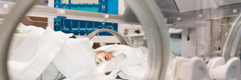
In neonatal intensive care, ventilating premature infants remains one of the greatest challenges. Their lungs are fragile and not yet fully developed, and synchronizing mechanical ventilation with the infant’s own breathing effort can be difficult.
In Banská Bystrica, Slovakia, clinicians applied NAVA with diaphragm monitoring (Edi) for the first time in 2021 in a premature infant with severe bronchopulmonary dysplasia (BPD). The baby’s condition improved, and before transfer, she was on spontaneous ventilation. Since then, the team has treated 34 infants with NAVA and continued using the Edi catheter beyond the conventional transition period, as the monitoring provided valuable information. (Read more)